Product Description
CAV3 Antibody | 31-074 | ProSci
Host: Rabbit
Reactivity: Human, Dog
Homology: N/A
Immunogen: Antibody produced in rabbits immunized with a synthetic peptide corresponding a region of human CAV3.
Research Area: Membrane
Tested Application: E, WB
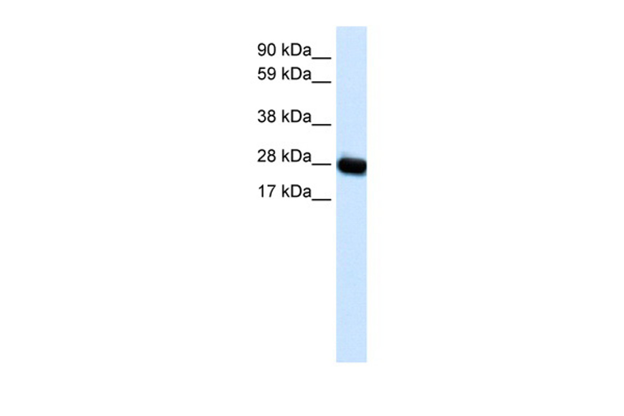
Application: CAV3 antibody can be used for detection of CAV3 by ELISA at 1:12500. CAV3 antibody can be used for detection of CAV3 by western blot at 0.5 μg/mL, and HRP conjugated secondary antibody should be diluted 1:50, 000 - 100, 000.
Specificiy: N/A
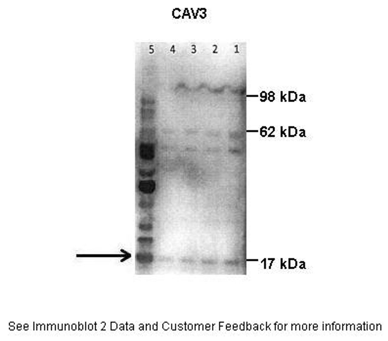
Positive Control 1: Cat. No. XBL-10413 - Fetal Skeletal Muscle Tissue Lysate
Positive Control 2: N/A
Positive Control 3: N/A
Positive Control 4: N/A
Positive Control 5: N/A
Positive Control 6: N/A
Molecular Weight: 17 kDa
Validation: N/A
Isoform: N/A
Purification: Antibody is purified by peptide affinity chromatography method.
Clonality: Polyclonal
Clone: N/A
Isotype: N/A
Conjugate: Unconjugated
Physical State: Liquid
Buffer: Purified antibody supplied in 1x PBS buffer with 0.09% (w/v) sodium azide and 2% sucrose.
Concentration: batch dependent
Storage Condition: For short periods of storage (days) store at 4˚C. For longer periods of storage, store CAV3 antibody at -20˚C. As with any antibody avoid repeat freeze-thaw cycles.
Alternate Name: CAV3, LQT9, VIP21, LGMD1C, VIP-21
User Note: Optimal dilutions for each application to be determined by the researcher.
BACKGROUND: CAV3 is a caveolin family member, which functions as a component of the caveolae plasma membranes found in most cell types. Caveolin proteins are proposed to be scaffolding proteins for organizing and concentrating certain caveolin-interacting molecules. Mutations identified in this gene lead to interference with protein oligomerization or intra-cellular routing, disrupting caveolae formation and resulting in Limb-Girdle muscular dystrophy type-1C (LGMD-1C) , hyperCKemia or rippling muscle disease (RMD) . Alternative splicing has been identified for this locus, with inclusion or exclusion of a differentially spliced intron. In addition, transcripts utilize multiple polyA sites and contain two potential translation initiation sites.
 Euro
Euro
 USD
USD
 British Pound
 NULL
NULL


