Product Description
IPO-38 Antibody [IPO-38] | 33-527 | ProSci
Host: Mouse
Reactivity: Human, Mouse, Rat
Homology: N/A
Immunogen: Spleen cells from a patient with hairy cell leukemia were used as the immunogen for the IPO-38 antibody.
Research Area: Cancer
Tested Application: Flow, IF, IHC-P
Application: Flow Cytometry: 0.5-1 ug/million cells in 0.1ml
Immunofluorescence: 0.5-1 ug/ml
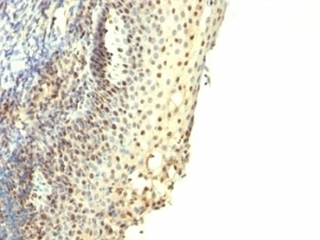
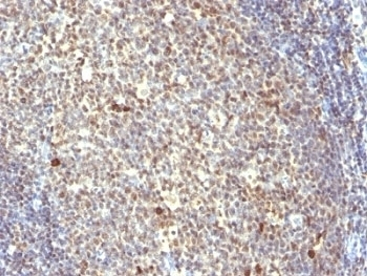
Immunohistochemistry (FFPE) : 0.5-1 ug/ml for 30 min at RT (1)
Prediluted format: incubate for 30 min at RT (2)
Optimal dilution of the IPO-38 antibody should be determined by the researcher.
1. Staining of formalin-fixed tissues requires boiling tissue sections in 10mM Citrate buffer, pH 6.0, for 10-20 min followed by cooling at RT for 20 min.
2. The prediluted format is supplied in a dropper bottle and is optimized for use in IHC. After epitope retrieval step (if required) , drip mAb solution onto the tissue section and incubate at RT for 30 min.
Specificiy: N/A
Positive Control 1: N/A
Positive Control 2: N/A
Positive Control 3: N/A
Positive Control 4: N/A
Positive Control 5: N/A
Positive Control 6: N/A
Molecular Weight: N/A
Validation: N/A
Isoform: N/A
Purification: PEG precipitation
Clonality: Monoclonal
Clone: IPO-38
Isotype: IgM, kappa
Conjugate: Unconjugated
Physical State: Liquid
Buffer: PBS with 0.1 mg/ml BSA and 0.05% sodium azide
Concentration: 0.2 mg/mL
Storage Condition: Aliquot and Store at 2-8˚C. Avoid freez-thaw cycles.
Alternate Name: N/A
User Note: Optimal dilutions for each application to be determined by the researcher
BACKGROUND: Recognizes a protein of 14-16kDa, which is a novel nuclear antigen of proliferating cells. IPO38 antigen is present in the nuclei of proliferating cells such as Hodgkin s disease and non-Hodgkin s lymphomas, different forms of leukemias, breast and colorectal carcinomas, and PHA-stimulated lymphocytes. It is not expressed in the cells of non-stimulated lymphocytes and granulocytes. IPO38 may be a useful marker of cell proliferation during monitoring of tumor progression.
 Euro
Euro
 USD
USD
 British Pound
 NULL
NULL

![IPO-38 Antibody [IPO-38] IPO-38 Antibody [IPO-38]](https://cdn11.bigcommerce.com/s-452hpg8iuh/images/stencil/1280x1280/products/575362/811967/porsci_lo__79508.1648973713__83221.1649091902.png?c=2)


![IPO-38 Antibody [IPO-38] IPO-38 Antibody [IPO-38]](https://cdn11.bigcommerce.com/s-452hpg8iuh/images/stencil/100x100/products/575362/811967/porsci_lo__79508.1648973713__83221.1649091902.png?c=2)


![IPO-38 Antibody [IPO-38] IPO-38 Antibody [IPO-38]](https://cdn11.bigcommerce.com/s-452hpg8iuh/images/stencil/500x659/products/575362/811967/porsci_lo__79508.1648973713__83221.1649091902.png?c=2)


