Product Description
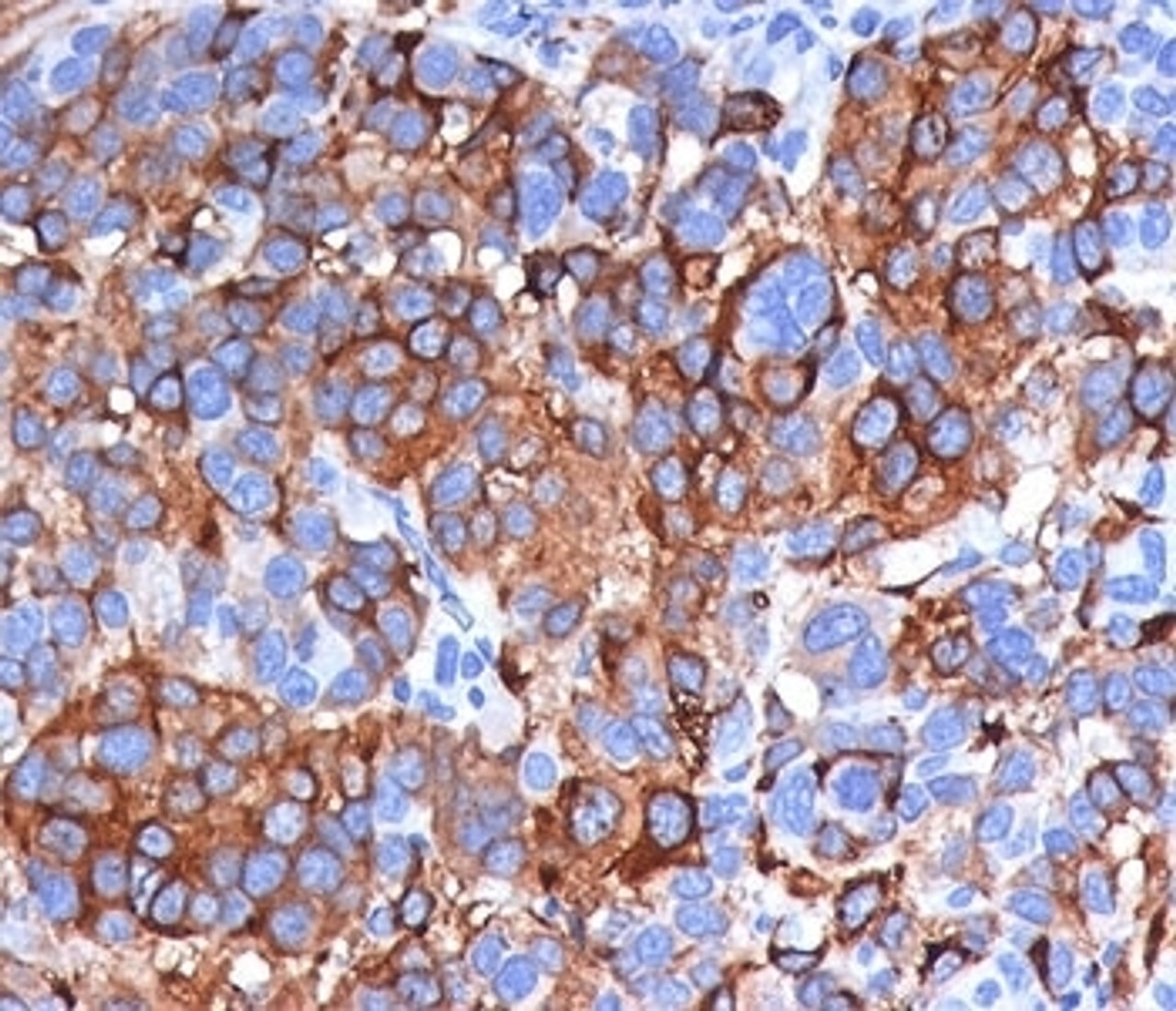
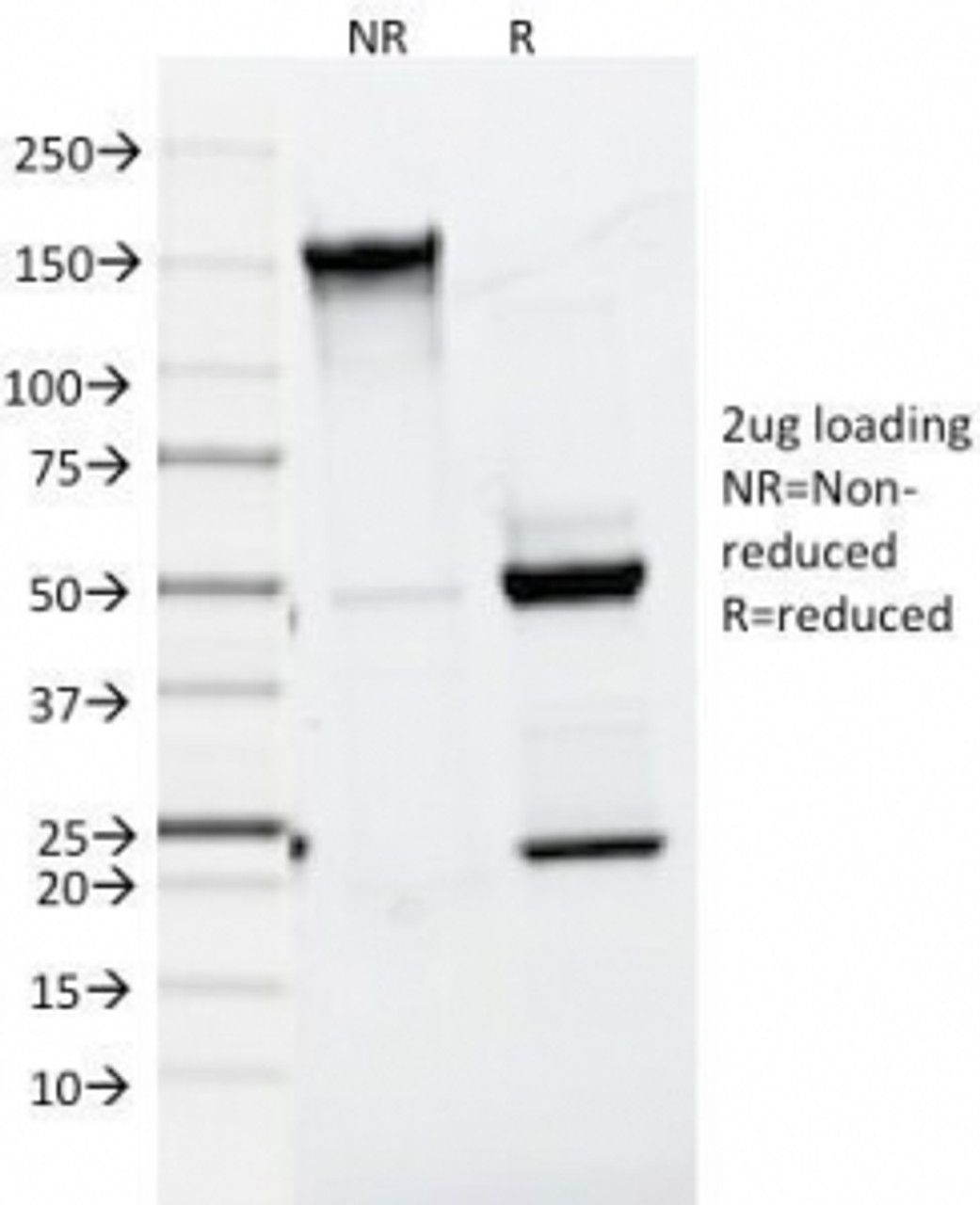
Melan-A Antibody [A103] | 33-132 | ProSci
Host: Mouse
Reactivity: Human, Mouse, Rat, Dog
Homology: N/A
Immunogen: Recombinant human protein was used as the immunogen for the Melan-A antibody.
Research Area: Immunology
Tested Application: WB, Flow, IHC, IF
Application: Flow Cytometry: 0.5-1 ug/million cells
IF: 0.5-1 ug/ml
WB: 0.5-1.0 ug/ml
IHC (FFPE) : 0.5-1 ug/ml for 30 minutes at RT (1)
Prediluted format : incubate for 30 min at RT (2)
Due to differences in protocols and secondary antibody used, the Melan-A antibody may require titration for optimal performance.
1. FFPE staining is enhanced by boiling sections in 10mM Citrate Buffer, pH 6.0, for 10-20 min followed by cooling at RT for 20 minutes.
2. The prediluted format is supplied in a dropper bottle and is optimized for use in IHC. After epitope retrieval step (if required) , drip mAb solution onto the tissue section and incubate at RT for 30 min.
Specificiy: N/A
Positive Control 1: N/A
Positive Control 2: N/A
Positive Control 3: N/A
Positive Control 4: N/A
Positive Control 5: N/A
Positive Control 6: N/A
Molecular Weight: N/A
Validation: N/A
Isoform: N/A
Purification: Protein G affinity chromatography
Clonality: Monoclonal
Clone: A103
Isotype: IgG1, kappa
Conjugate: Unconjugated
Physical State: Liquid
Buffer: PBS with 0.1 mg/ml BSA and 0.05% sodium azide
Concentration: 0.2 mg/mL
Storage Condition: Aliquot and Store at 2-8˚C. Avoid freez-thaw cycles.
Alternate Name: MLANA, Antigen LB39-AA, Antigen SK29-AA, Melan-A, Protein Melan-A, MART-1, MART1
User Note: Optimal dilutions for each application to be determined by the researcher
BACKGROUND: Antibody A103 is specific for Melan-A (likely an abbreviation for ‘melanocyte antigen’) , also called MART-1, a unique antigen on melanoma cells recognized by CTLs (cytotoxic T lymphocytes) , making it useful as a melanocyte differentiation marker. It is commonly used to identify metastatic tumors of melanocytic origin, as well as perivascular epithelioid cell tumors. Unlike another commonly used Melan-A / MART-1 antibody, M2-7C10, A103 also stains steroid hormone producing cells, making it valuable in identifying adrenocortical carcinomas.
Note: this protein was identified in 1994 by two different labs, one naming it MART-1 (melanoma antigen recognized by T-cells) and the other Melan-A. Both names are commonly used in literature, but clone A103 antibody is generally referred to as being anti-Melan-A.
 Euro
Euro
 USD
USD
 British Pound
 NULL
NULL

![Melan-A Antibody [A103] Melan-A Antibody [A103]](https://cdn11.bigcommerce.com/s-452hpg8iuh/images/stencil/1280x1280/products/575000/811135/porsci_lo__79508.1648973713__10708.1649091782.png?c=2)


![Melan-A Antibody [A103] Melan-A Antibody [A103]](https://cdn11.bigcommerce.com/s-452hpg8iuh/images/stencil/100x100/products/575000/811135/porsci_lo__79508.1648973713__10708.1649091782.png?c=2)


![Melan-A Antibody [A103] Melan-A Antibody [A103]](https://cdn11.bigcommerce.com/s-452hpg8iuh/images/stencil/500x659/products/575000/811135/porsci_lo__79508.1648973713__10708.1649091782.png?c=2)


![Melan-A Antibody [MRT3V] Melan-A Antibody [MRT3V]](https://cdn11.bigcommerce.com/s-452hpg8iuh/images/stencil/500x659/products/575663/812723/porsci_lo__79508.1648973713__19418.1649092001.png?c=2)

