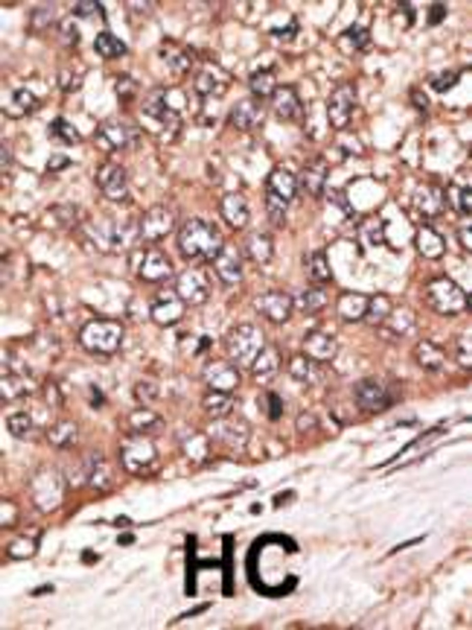
Product Description
WISP1 Antibody | 62-199 | ProSci
Host: Rabbit
Reactivity: Human
Homology: N/A
Immunogen: This WISP1 antibody is generated from rabbits immunized with a KLH conjugated synthetic peptide between 171-200 amino acids from the Central region of human WISP1.
Research Area: Signal Transduction
Tested Application: WB, IHC-P
Application: For WB starting dilution is: 1:1000
For IHC-P starting dilution is: 1:50~100
Specificiy: N/A
Positive Control 1: N/A
Positive Control 2: N/A
Positive Control 3: N/A
Positive Control 4: N/A
Positive Control 5: N/A
Positive Control 6: N/A
Molecular Weight: 40 kDa
Validation: N/A
Isoform: N/A
Purification: This antibody is prepared by Saturated Ammonium Sulfate (SAS) precipitation followed by dialysis
Clonality: Polyclonal
Clone: N/A
Isotype: Rabbit Ig
Conjugate: Unconjugated
Physical State: Liquid
Buffer: Supplied in PBS with 0.09% (W/V) sodium azide.
Concentration: batch dependent
Storage Condition: Store at 4˚C for three months and -20˚C, stable for up to one year. As with all antibodies care should be taken to avoid repeated freeze thaw cycles. Antibodies should not be exposed to prolonged high temperatures.
Alternate Name: WNT1-inducible-signaling pathway protein 1, WISP-1, CCN family member 4, Wnt-1-induced secreted protein, WISP1, CCN4
User Note: Optimal dilutions for each application to be determined by the researcher.
BACKGROUND: Wisp1 is a member of the WNT1 inducible signaling pathway (WISP) protein subfamily, which belongs to the connective tissue growth factor (CTGF) family. WNT1 is a member of a family of cysteine-rich, glycosylated signaling proteins that mediate diverse developmental processes. The CTGF family members are characterized by four conserved cysteine-rich domains: insulin-like growth factor-binding domain, von Willebrand factor type C module, thrombospondin domain and C-terminal cystine knot-like domain. Wisp1 may be downstream in the WNT1 signaling pathway that is relevant to malignant transformation. It is expressed at a high level in fibroblast cells, and overexpressed in colon tumors. The encoded protein binds to decorin and biglycan, two members of a family of small leucine-rich proteoglycans present in the extracellular matrix of connective tissue, and possibly prevents the inhibitory activity of decorin and biglycan in tumor cell proliferation. It also attenuates p53-mediated apoptosis in response to DNA damage through activation of the Akt kinase. It is 83% identical to the mouse protein at the amino acid level.
 Euro
Euro
 USD
USD
 British Pound
 NULL
NULL


