Product Description
NOX3 Antibody | 7925 | ProSci
Host: Rabbit
Reactivity: Human, Mouse
Homology: N/A
Immunogen: NOX3 antibody was raised against a 15 amino acid peptide near the amino terminus of human NOX3.
The immunogen is located within amino acids 110 - 160 of NOX3.
Research Area: Stem Cell
Tested Application: E, WB, IHC-P, IF
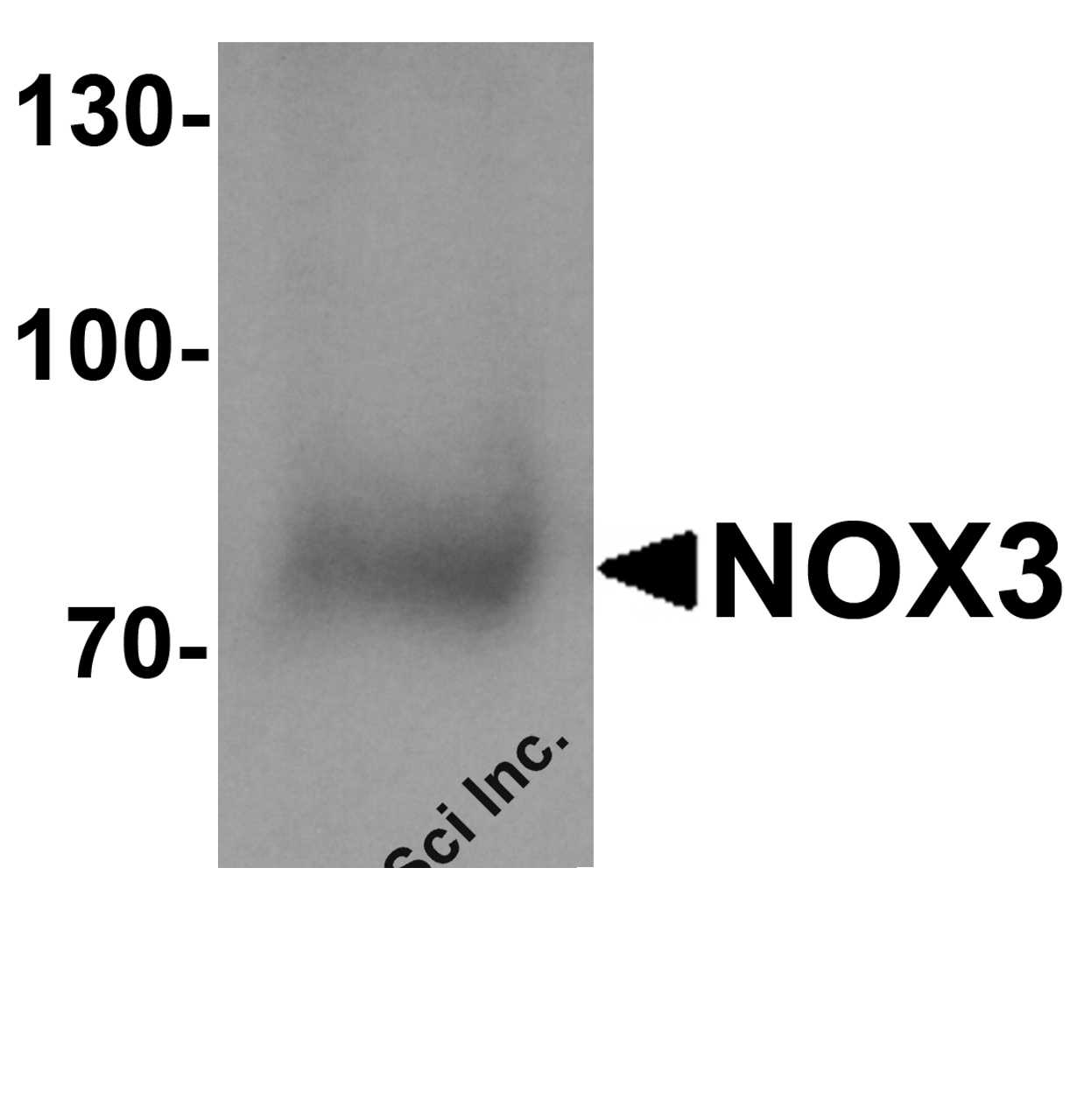
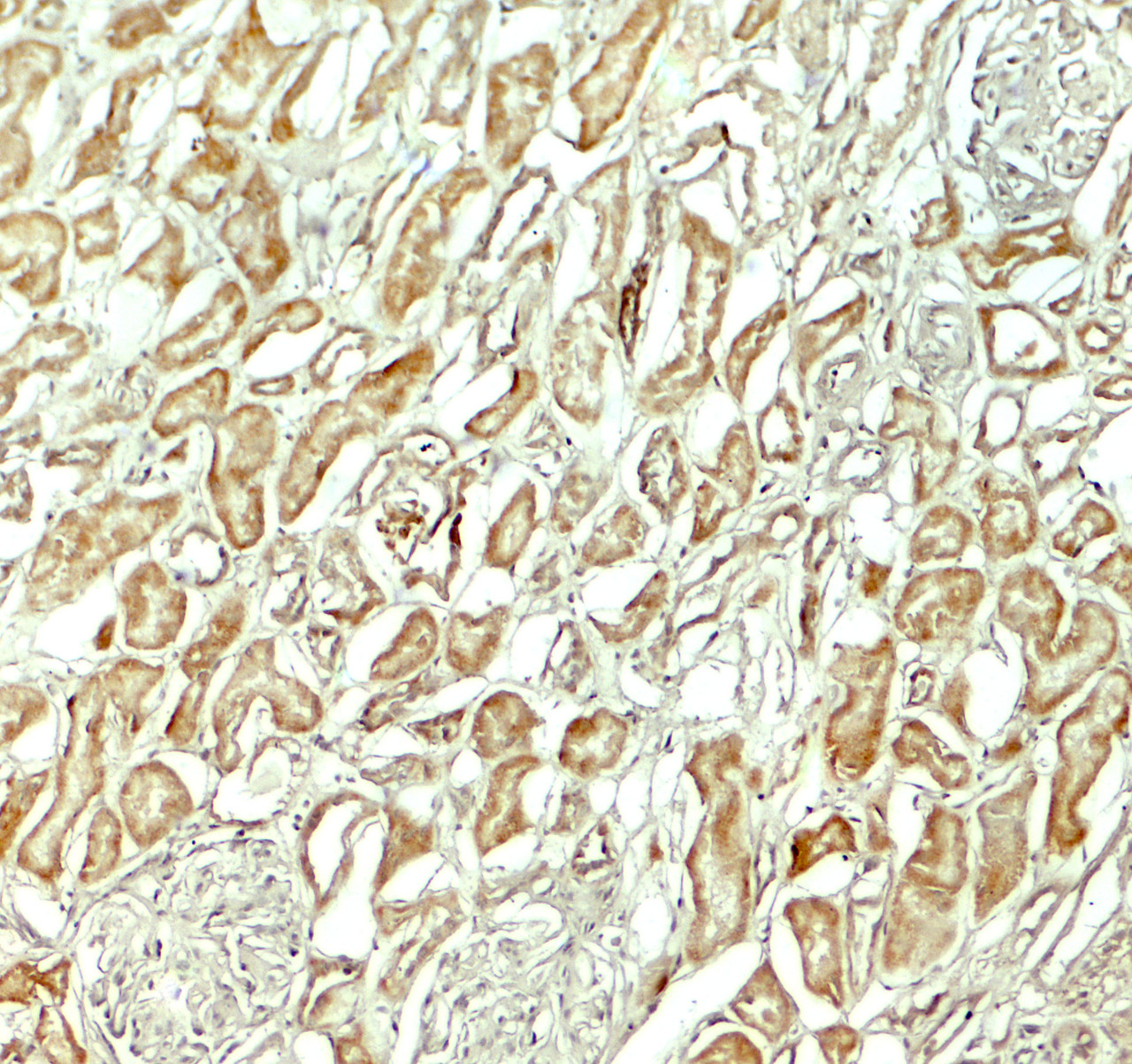
Application: NOX3 antibody can be used for detection of NOX3 by Western blot at 1 - 2 μg/ml. Antibody can also be used for Immunohistochemistry starting at 5 μg/mL. For immunofluorescence start at 20 μg/mL.
Antibody validated: Western Blot in human samples; Immunohistochemistry in human samples and Immunofluorescence in human samples. All other applications and species not yet tested.
Specificiy: NOX3 antibody is human and mouse reactive. NOX3 is predicted to not cross-react with other NOX proteins.
Positive Control 1: Cat. No. 1210 - HEK293 Cell Lysate
Positive Control 2: Cat. No. 10-401 - Human Kidney Tissue Slide
Positive Control 3: N/A
Positive Control 4: N/A
Positive Control 5: N/A
Positive Control 6: N/A
Molecular Weight: Predicted: 62 kDa
Observed: 72 kDa
Validation: N/A
Isoform: N/A
Purification: NOX3 antibody is affinity chromatography purified via peptide column.
Clonality: Polyclonal
Clone: N/A
Isotype: IgG
Conjugate: Unconjugated
Physical State: Liquid
Buffer: NOX3 antibody is supplied in PBS containing 0.02% sodium azide.
Concentration: 1 mg/mL
Storage Condition: NOX3 antibody can be stored at 4˚C for three months and -20˚C, stable for up to one year.
Alternate Name: NAPDH oxidase 3, NOX-3, GP91-3, MOX2, MOX-2
User Note: Optimal dilutions for each application to be determined by the researcher.
BACKGROUND: The NOX family of NAPDH oxidases is comprised of seven transmembrane proteins that oxidize intracellular NAPDH/NADH, causing electron transport across the membrane and the reduction of molecular oxygen to superoxide (1) . NOX3 is expressed predominantly in the inner ear and is involved in the biogenesis of otoconia/otolith (2, 3) . It has been suggested that NOX3 is activated by the Transient Receptor Potential Vanilloid 1 (TRPV1) , and this activity causes increased levels of reactive oxygen species in the inner ear, which in turn leads to STAT1-mediated inflammation and hearing loss (4) .
 Euro
Euro
 USD
USD
 British Pound
 NULL
NULL


